X-Ray Micro-Computed Tomography (Micro-CT)
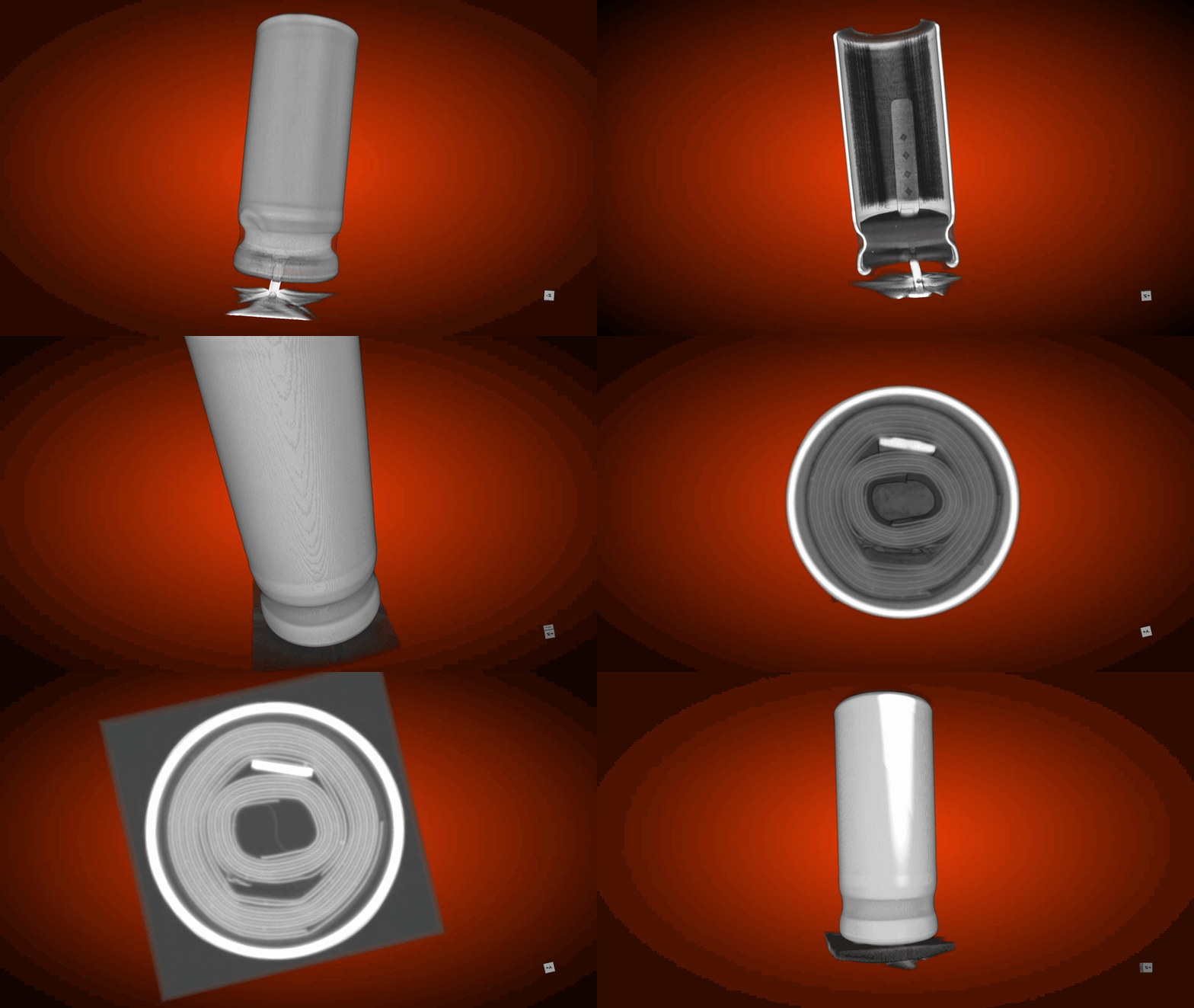
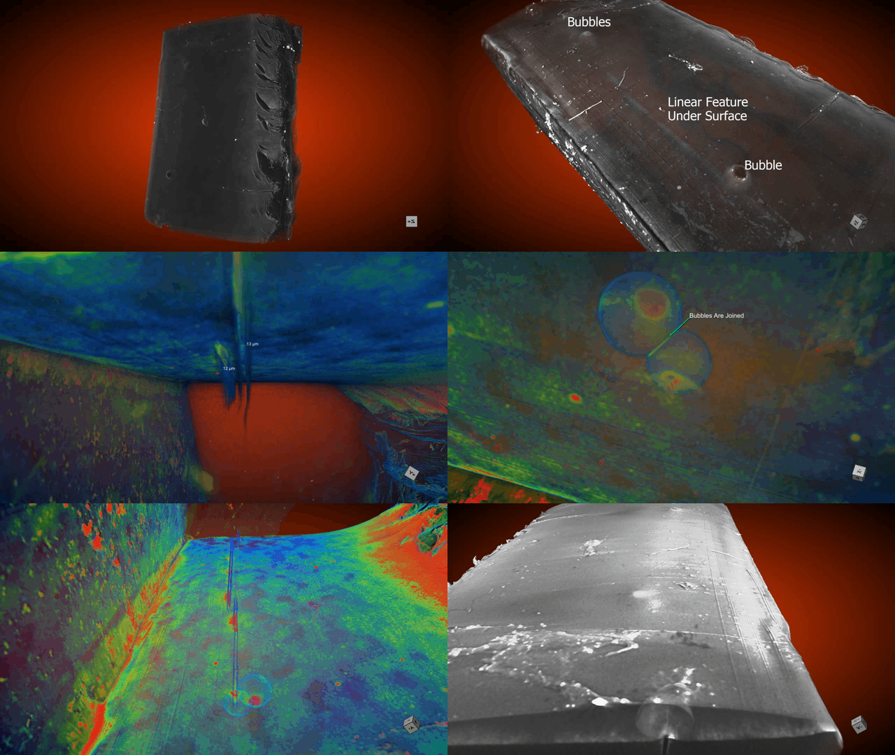
Similar to a hospital CAT scanning system, Micro Computed Tomography (Micro-CT) is a non-destructive 3D imaging technique (4-D with time-based measurements) that uses a highly energetic X-Ray beam to create a series of 2D projections, whose greyscales vary with the volume’s internal density and atomic number variations. These projections are reconstructed to create a virtual 3D model, allowing users to “see” inside the sample without preparing, sectioning or destroying the sample.
Instrument:
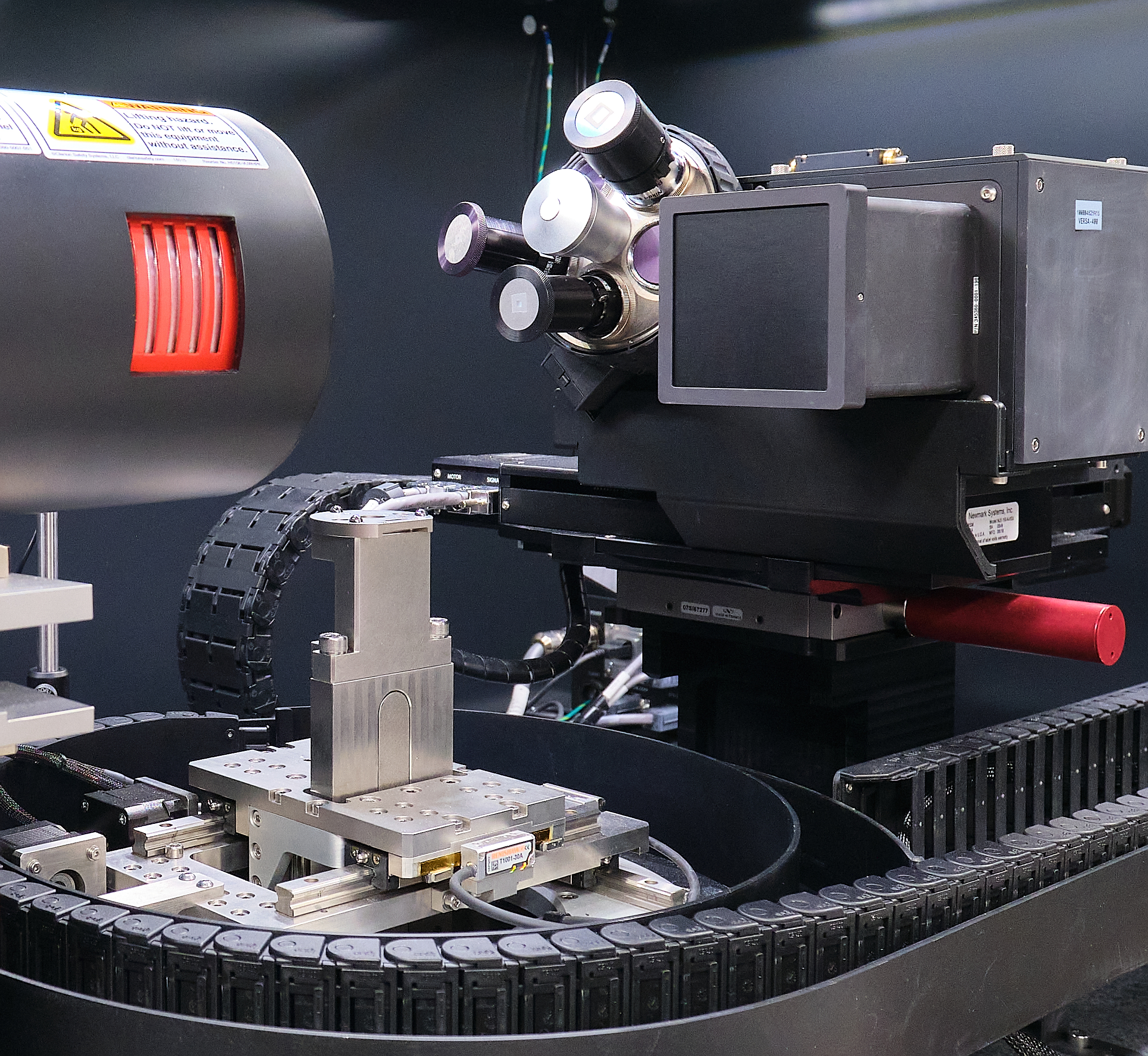
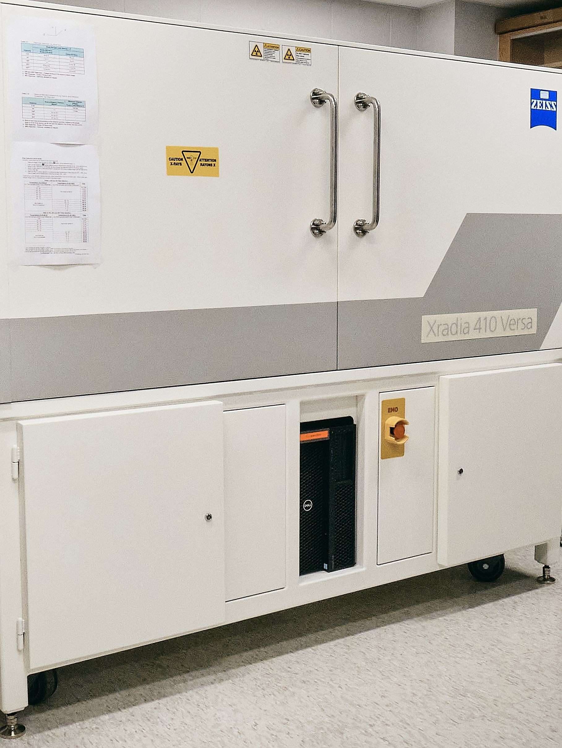
Zeiss Xradia 410 Versa Micro-CT
System Capabilities:
- Minimum spatial resolution of 0.9 µm and a minimum voxel size of 100 nm at larger working distances
- Variable X-Ray voltage from 0 to 150 kV
- Variable X-Ray power from 0 to 10 W
- Automated objective carousel, with objectives: 0.4x, 4x, 10x, 20x
- Advanced absorption and phase contrast capability (for soft or low atomic number materials) overcomes the limitations on traditional computer tomography
- Vertical stitching mode enables the analysis of taller samples by joining tomographies from different sections of the sample
- The in-situ tensile stage (5000 N) enables measurements while the samples are under tensile or compression stresses
Selected Applications in Industry:
- Corrosion analysis within an alloy
- Distribution of porosities in metals, oil reservoir rocks, and other samples
- Phase contrast analysis of internal phases
- Failure analysis of large intact circuit boards and other 3-D packages
- Non-destructive reverse engineering of obsolete Integrated Circuit packages
- Defects in polymers and paint coatings
- Imaging/mapping of biological, pathological, and neural networks