The use of low duty cycle pulsed-unipolar current mode for producing Alumina/ZnO nanocomposite coatings via plasma electrolytic oxidation process, S. Shahzamani, A. Ashrafi, M.R. Toroghinejad, V. Dehnavi and A. Hakimizad, Materials Research Express (2019), 6, 076555. Read Full Text
Galvanic interaction of pyrite with Cu activated sphalerite and its effect on xanthate adsorption, L. Xia, B. Hart, F. Larachi and O. Gravel, The Canadian Journal of Chemical Engineering (2019), 97, 2671-2677. Read Full Text
Correlation between the hydrogen peroxide formed during grinding and the oxidized species present on the surface of sphalerite, L. Xia and B.R. Hart, Minerals Engineering (2019), 130, 165-170. Read Full Text

Researchers at Surface Science Western, Western University and Mawson Institute, University of South Australia have developed a variety of X-ray photoelectron spectroscopy (XPS) protocols that allow for a fuller understanding of transition metal chemistries at surfaces. Recent work, published in Physical Chemistry Chemical Physics, provides an in-depth look at the bonding within nickel-containing alloys, oxides and halides. Previous work on nickel surfaces has allowed for semi-quantitative analysis of the nickel species present in a sample. As well, a more accurate calculation for estimating oxide film thicknesses on a nickel metal surface has been developed.
These techniques have been expanded upon to cover the entire set of first row transition metals (see publications for (Cr), (Fe), (Ni), (Sc, Ti, V, Cu and Zn) and (Cr, Mn, Fe, Co and Ni)).
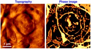
Researchers at Surface Science Western and the Kilee Patchell-Evans Autism Research Group at Western University have developed an atomic force microscope (AFM) phase imaging technique that allows for imaging of subcellular structures in unfixed rat brain sections. The contrast in phase images originates from the difference in mechanical properties between biological structures. Visualization of the native state of biological structures by way of their mechanical properties provides a complementary technique to more traditional imaging techniques such as optical and electron microscopy. The results have been published (PDF) in volume 136 (2011) of the Analyst.

Researchers at the Chinese University of Hong Kong and Surface Science Western have demonstrated a simple, convenient, nonaqueous electrochemical approach to fabricating various single-crystal chitosan nanostructures. This approach does not require a template, catalysis, or surfactant. The nanostructures are grown by optimal deposition time, electrochemical potential, and the electric current. The method provides a variety of nanomorphologies for further applications and studies, for example, cell adhesion, in vitro cell proliferation, tissue-compatible scaffolds, and nanostructure-based sensors. Considering the proven versatility and biocompatibility of chitosan, one may find profound applications of chitosan nanowires in nanodevices, particularly in the medical and pharmaceutical fields. The results have been published (PDF) in the June 4, 2008 issue of Advance Materials.